Introduction
Foot pathologies
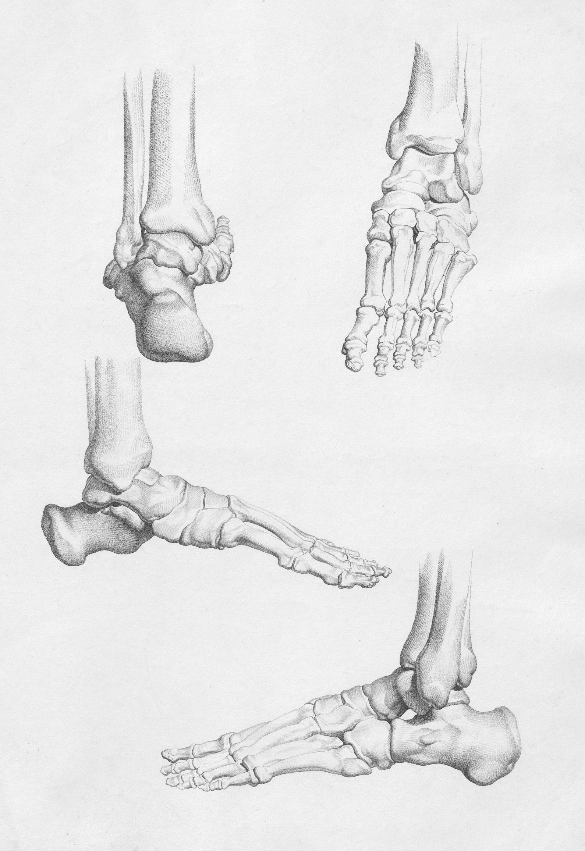
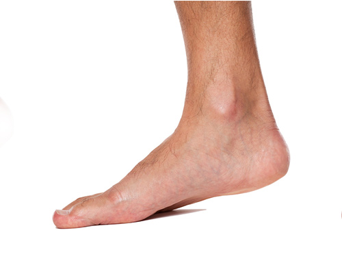
The foot is a complex structure of 26 bones, essential for balance and walking. Hallux valgus, metatarsalgia, plantar fasciitis and post-traumatic pathologies require accurate biomechanical evaluation.
Techniques
Surgical and conservative techniques
Mini-invasive microsurgery, corrective osteotomies, custom orthotics, biological therapies for early cartilage wear.
Foot anatomy
The foot is an extremely complex structure representing the most distal part of the lower limb: 26 bones surrounded by cartilaginous tissue that ensures fluid movement. Any trauma damaging the internal tissue can compromise its stability. Foot surgery has undergone extraordinary development over recent decades, offering therapeutic solutions previously unimaginable. Prof. Colao brings thirty years of experience with the most advanced diagnostic and microsurgical techniques.
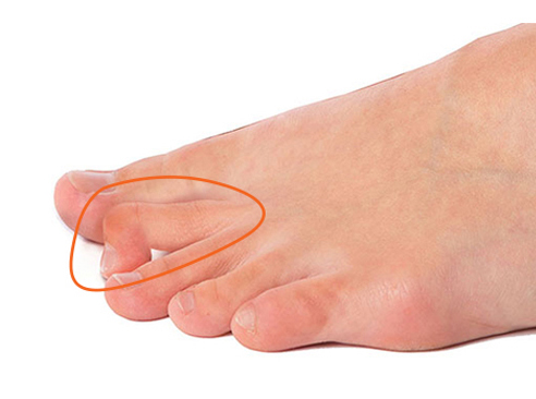
Hammer toes
Hammer toe is a deformity characterised by flexion of the interphalangeal joint and dorsal hyperextension of the metatarsophalangeal and distal interphalangeal joints: the toe takes the shape of a piano hammer. Beyond the aesthetic alteration, it causes persistent pain. Causes range from inappropriate footwear to genetic, traumatic or rheumatic factors. The short-duration procedure involves an osteotomy at the base of the phalanx or tenotomy of the flexors and/or extensors.
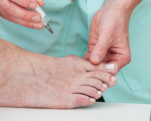
Hallux valgus
Hallux valgus correction is one of Prof. Colao's team's strengths: the microsurgery procedure ensures rapid recovery with no need for crutches or special footwear. The speed of gait recovery is remarkable. Microsurgery eliminates foot pain, allows return to all favourite sports and enables wearing any type of footwear without restrictions.
Flatfoot endorthesis
Surgical treatment of flatfoot in growing children aims to correct hyperpronation of the subtalar joint — between the calcaneus and talus — to allow normal development of the foot, legs and spine. The arthroscopic procedure restricts the articular glide between talus and calcaneus. For 7-8 days the joint is immobilised in a weight-bearing plaster boot; the child can walk immediately after. The implants are removed after 2-3 years.
Clinical information
Hammer toes
- Procedure
- Mini-invasive surgery (phalangeal osteotomy or tenotomy)
- Hospital stay
- Day hospital
- Anaesthesia
- Local anaesthesia at the operative site
- Post-op course
- Light activity resumed within a few days
- Rehabilitation
- No rehabilitation period required
Hallux valgus
- Procedure
- Micro-invasive surgery with percutaneous osteotomy
- Hospital stay
- Day hospital
- Anaesthesia
- Local anaesthesia at the operative site
- Post-op course
- Immediate walking post-op with specific footwear
- Rehabilitation
- No rehabilitation required (except in elderly patients)
Flatfoot endorthesis
- Procedure
- Subtalar arthroscopy with endorthesis implant
- Hospital stay
- Day hospital
- Anaesthesia
- Regional, general or combined anaesthesia
- Post-op course
- Weight-bearing plaster boot for 7-8 days; walking on completion
- Rehabilitation
- 2-3 months of check-ups; endorthesis removal after 2-3 years
The information has educational value and does not replace specialist medical examination.
Book a visit