Introduction
Lumbosacral pathologies
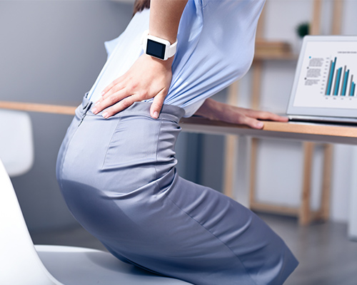
The lumbosacral region bears the highest load. Disc herniations, canal stenosis, mechanical low back pain and sciatica are assessed with a multidisciplinary approach.
Techniques
Surgical and conservative techniques
Imaging diagnostics, pharmacological and physical therapy, guided infiltrations, selective decompressive microsurgery.
Lumbosacral anatomy
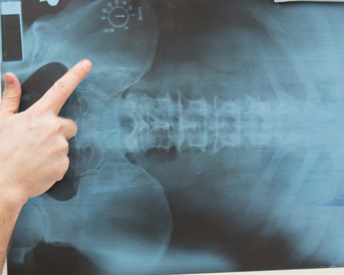
The human vertebral column consists of 33-34 vertebrae: 7 cervical, 12 thoracic, 5 lumbar, 5 sacral and 4-5 coccygeal, the latter fused into a single structure. The lumbosacral junction is where the fifth lumbar vertebra articulates with the upper sacrum. This region bears the greatest mechanical load: the lumbar vertebrae are the largest and strongest, while the sacrum forms the posterior wall of the pelvis. Intervertebral discs between each vertebral pair absorb loads and enable flexion, extension and rotation.

Lumbar disc herniation
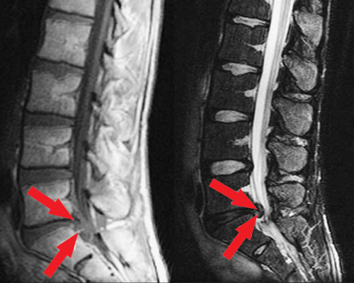
Disc herniation, also known as disc prolapse, consists in the rupture of the fibrous annulus of the intervertebral disc: the nucleus pulposus leaks out and compresses nearby nerve roots. Causes are multiple — mainly age-related degeneration, sudden exertion or prolonged incorrect posture. Symptoms include acute or chronic low back pain, sciatica, tingling and motor deficits in the lower limbs.
Vertebral disc disease
Disc disease is a degenerative condition affecting the intervertebral disc, compromising its structure and shock-absorbing function. The fibrous capsule deteriorates progressively, producing leakage of fluid from the nucleus pulposus. In most cases anti-inflammatory drug therapy promotes reabsorption and pain control. Pain arises from compression of nerve endings within the vertebral canal.
The information has educational value and does not replace specialist medical examination.
Book a visit